Digital X-Rays
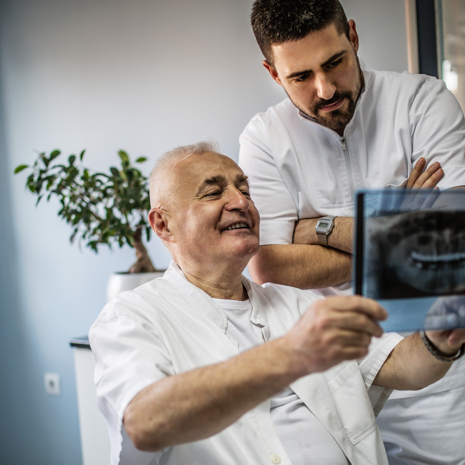
Digital radiographs (x-rays) are an important diagnostic tool for dental professionals. While traditional film radiographs provide critical insight into the oral and physical health of the patient, hi-tech digital radiographs allow dentists to view and enhance dental images on a large computer screen.
Dentists can also copy or print digital radiographs with ease. This allows for effective comparison of new results to previous images and insight on how treatments have impacted dental conditions. If the dentist refers the patient to a specialist, digital radiographs can be transmitted via computer – eliminating the need for a second set of x-rays in most cases.
Why Use Digital Radiographs?
One of the most significant advantages of utilizing digital radiographs is reduction of radiation exposure. Digital radiographs also eliminate the use of film and required chemicals for processing, making the overall procedure much less harmful to the environment.
The larger computer screen used to display digital radiographs allows dentists to view any problems or irregularities with added clarity. The potential for early detection of decay or periodontal problems and reducing complicated conditions later is vastly increased.
Here are some of the main conditions that digital radiographs can better expose:
- Small areas of decay
- Bone recession
- Tumors
- Fractures and trauma
- Positioning of the teeth
- Developmental irregularities
- Tooth positioning
How Are Digital Radiographs Taken?
The technique for capturing digital radiographs is similar to that of the traditional-style radiographs, but the digital variety uses a small electronic sensor to capture intraoral images, as opposed to film bitewings.
Generally, a full mouth series of digital x-rays includes eighteen different views of the teeth and underlying jawbone. The two standard views dentists use are: periapical and bitewing. The periapical view is used to inspect the root tips for decay, disease or damage, while the bitewing view allows for close inspection and measurement of the mandible and maxilla (upper and lower jawbones).
After exposure, the digital image is either transferred wirelessly to a computer, or the dentist takes the plate from the mouth, and scans it with a specialized reader. Processing traditional film can take up to five minutes, but a digital image takes mere seconds. Once the image is apparent on the screen, the contrast, color and brightness can be altered to be much clearer.
What Are The Different Types of Digital Radiographs?
There are two main kinds of dental digital X-rays. Intraoral ones are either bitewing or periapical X-rays. Bitewings require the patient to bite down on a specialized sensor that can take the images of a specific area of the mouth. These X-rays can assess the integrity and fit of restorations, and can show the internal structure of an entire tooth & signs of bone loss or abscess formation.
Extraoral ones have panoramics that need a machine that rotates around the patient’s head. Panoramics provide one image of all the patient’s teeth in the lower and upper arches. These are effective in assessing jaw and dental problems. Because of these diagnostic exams, dentists could formulate more accurate treatment plans.
Are Digital
X-Rays Safe?
We are all exposed to natural radiation in our environment. The amount of radiation exposure from a full mouth series of X-rays is equal to the amount a person receives in a single day from natural sources.
Dental X-rays produce a low level of radiation and are considered safe. Dentists take necessary precautions to limit the patient’s exposure to radiation when taking dental X-rays. These precautions include using lead apron shields to protect the body and using modern, fast film that cuts down the exposure time of each X-ray.
How Often Should Dental
X-rays be Taken?
The need for dental X-rays depends on each patient’s individual dental health needs. Your dentist and dental hygienist will recommend necessary x-rays based on the review of your medical and dental history, dental exam, signs and symptoms, age consideration, and risk for disease.
A full mouth series of dental X-rays is recommended for new patients. A full series is usually good for three to five years. Bite-wing X-rays (X-rays of top and bottom teeth biting together) are taken at recall (check-up) visits and are recommended once or twice a year to detect new dental problems.
Dental X-rays
May Reveal:
- Abscesses or cysts
- Bone loss
- Cancerous and non-cancerous tumors
- Decay between the teeth
- Developmental abnormalities
- Poor tooth and root positions
- Problems inside a tooth
- Problems below the gum line
Detecting and treating dental problems at an early stage can save you time, money, unnecessary discomfort, and, of course, your teeth!